tree in bud radiology assistant
As the technique calls for 1-2 mm slice. This pattern is manifested by luminal filling of contiguous branching segments of bronchioles seen in bronchiolar disease.
Chronic Airspace Disease Review Of The Causes And Key Computed Tomography Findings
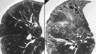
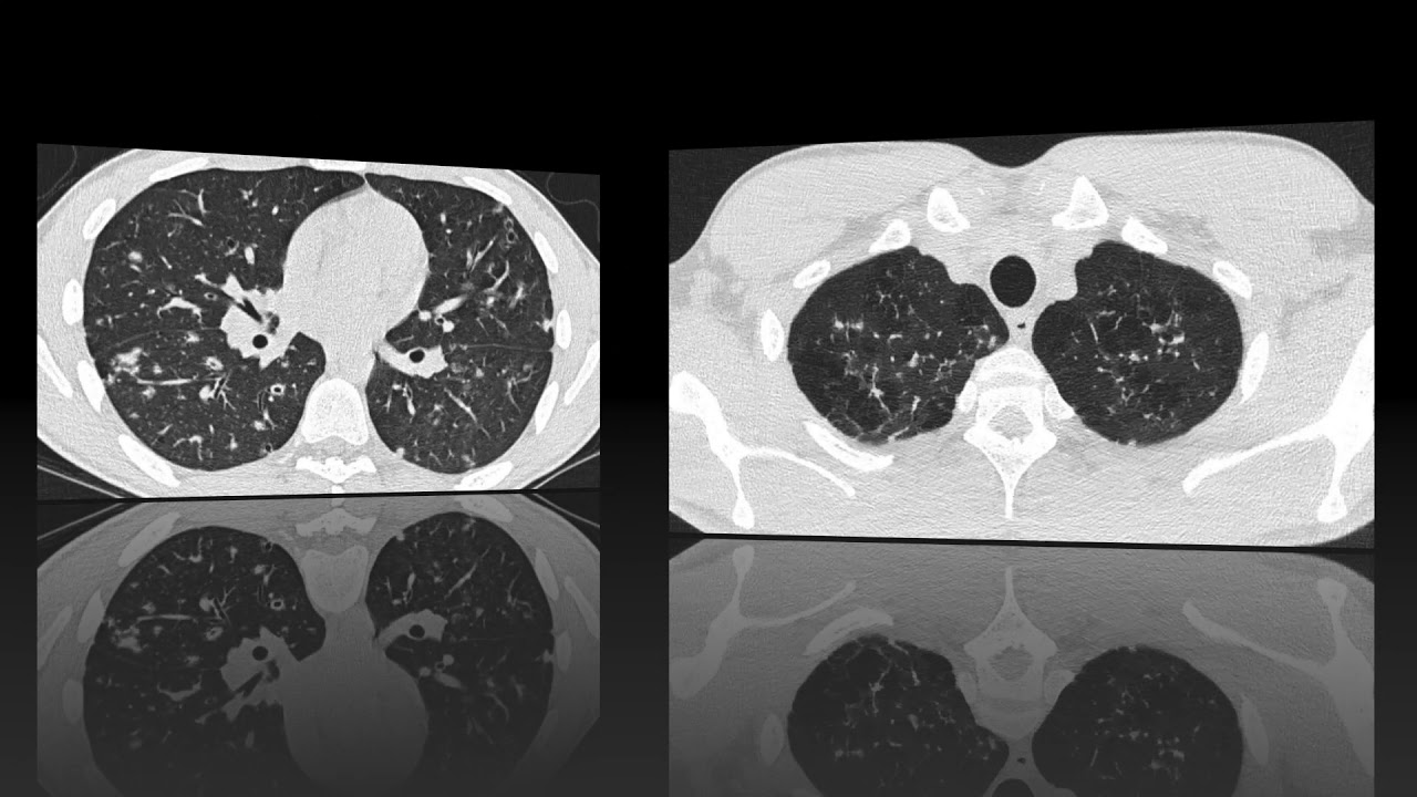
Abnormal tree-in-bud bronchioles can be distinguished from normal centrilobular bronchioles by their more irregular appearance lack of tapering or knobbybulbous appearance at the tip of their branches.
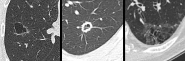
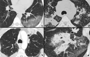
. It consists of small centrilobular nodules of soft-tissue attenuation connected to multiple branching linear structures of similar caliber that originate from a single stalk. Originally reported in cases of endobronchial spread of. In centrilobular nodules the recognition of tree-in-bud is of value for narrowing the differential diagnosis.
2-3 mm nodules with random disrtibution. However in some cases nodules occurring in relation to centrilobular arteries may mimic the appearance of the tree-in-bud pattern. Discover and save your own Pins on Pinterest.
Diagnosis Pathophys Radiology Pulmonary CTChest TreeInBud Diagram RadiologyAssistant. The tree-in-bud distribution is often. This pattern is most pronounced in the lung periphery and is usually associated with abnormalities of the larger.
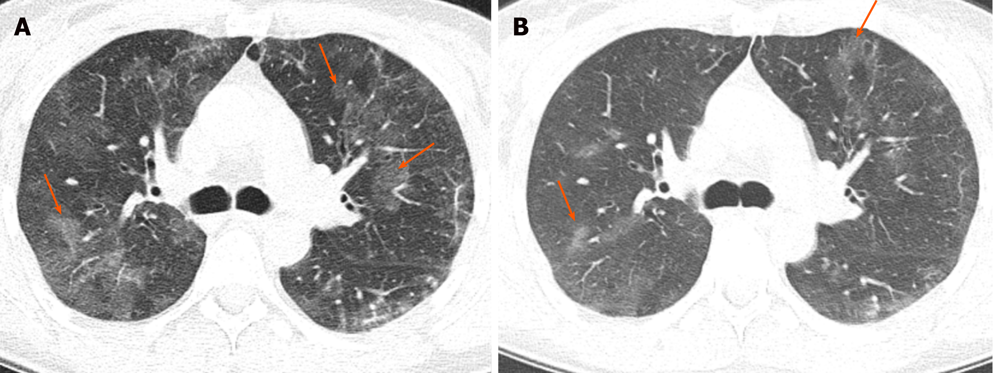
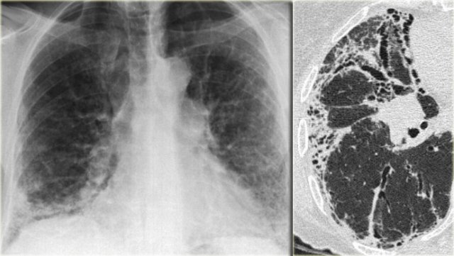
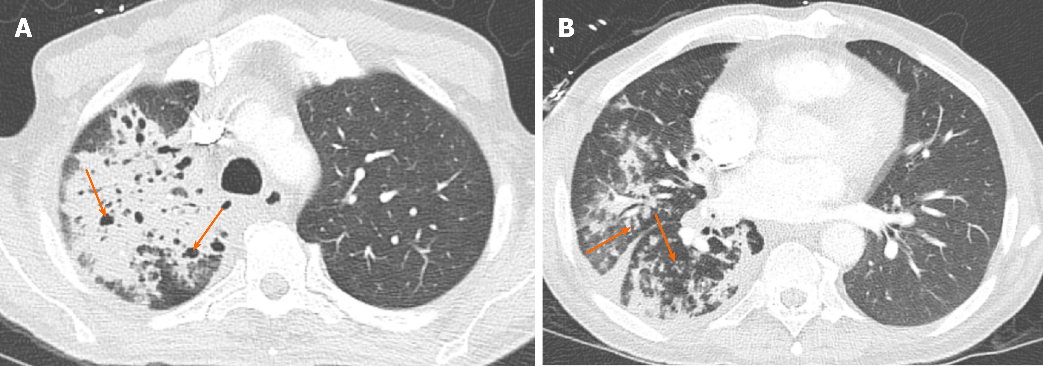
There is a cavitating lesion and typical tree-in-bud appearance. To describe the appearance of the endobronchial spread of mycobacterial tuberculosis. The blue arrow indicates the biopsy needle.
Mar 8 2020 - This Pin was discovered by Pankaj Kaira. It represents dilated and impacted mucus or pus-filled centrilobular bronchioles. Endobronchial spread of infection TB MAC any bacterial bronchopneumonia Airway disease associated with infection cystic fibrosis bronchiectasis less often an airway disease associated primarily with mucus retention allergic bronchopulmonary aspergillosis asthma.
We wish to emphasize that both clinicians and radiologists should be aware of the wide spectrum of disorders that can result in the tree-in-bud pattern. Oak Brook IL 60523-2251. The pattern reflects a spectrum of endo- and peribronchiolar disorders including mucoid impaction inflammation andor fibrosis 154.
Centrilobular nodularity and tree in bud appearances are not pathognomonic they are distinctive enough to strongly suggest TB910Presence of lymphadenopathy calcified or otherwise consolidation cavitation and the presence of pleural effusion are also helpful1 Good as HRCT is at depicting lung pathology it has its limitations. Usually somewhat nodular in appearance the tree-in-bud pattern is generally most pronounced in the lung periphery and associated with abnormalities of the larger airways. Tree in bud appearance.
Tree-in-bud almost always indicates the presence of. It represents dilated and impacted mucus or pus-filled centrilobular bronchioles. As you can see the possible causes of a tree in bud appearance are legion.
Tree-in-bud describes the appearance of an irregular and often nodular branching structure most easily identified in the lung periphery. Normal lobular bronchioles 1 mm in diameter cannot be seen on CT scans which can only show bronchi more than 2 mm in diameter. Bronchial wall thicken-ing is a potentially reversible finding and correlates with patient-reported symptoms health status and frequency of exacerbation 911.
Tree-in-bud In centrilobular nodules the recognition of tree-in-bud is of value for narrowing the differential diagnosis. Tree-in-bud describes the appearance of an irregular and often nodular branching structure most easily identified in the lung periphery. 820 Jorie Blvd Suite 200.
The goal of the classification system is to standardize follow-up and management decisions. Pus mucus or inflammatory exudate centrilobular bronchioles. Tree-in-bud TIB opacities are a common imaging finding on thoracic CT scan.
Where there is small airways disease and tree in bud is present this can be termed an exudative bronchiolitis. Tree-in-bud describes the appearance of an irregular and often nodular branching structure most easily identified in the lung periphery. In panbronchiolitis of East Asia particularly Japan a typical bilaterally symmetrical tree-in-bud pattern with lower lobe predominance is seen.
Occasionally tree-in-bud pattern may result from abnormalities of the centrilobular pulmonary arteries due to intravascular. Bronchiolitis and bronchiolectasis are nonspecific inflammatory. The tree-in-bud pattern is commonly seen at thin-section computed tomography CT of the lungs.
These findings serve as indirect signs and can increase the radiologists confidence in diag-nosing mild bronchiectasis. The tree-in-bud pattern was first used as a descriptor by Im et al. Tree-in-bud appearance represents dilated and fluid-filled ie.
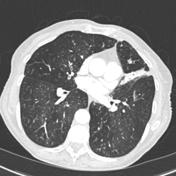
The tree-in-bud pattern represents centrilobular branching structures that resemble a budding tree. On the left a patient with TB. Lung-RADS or lung imaging reporting and data system is a classification proposed to aid with findings in low-dose CT screening exams for lung cancer.
Tree-in-bud pattern involving the dependent regions suggest aspiration bronchiolitis. The role of the radiologist is to narrow the list of differential diagnoses and to guide in. 1-4Reported causes include infections aspiration and a variety of infl ammatory conditions.
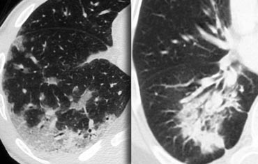
Plugging and tree-in-bud opacities figure 1c and table 1. However diseased bronchioles can be seen. Assistant Professor Department of.
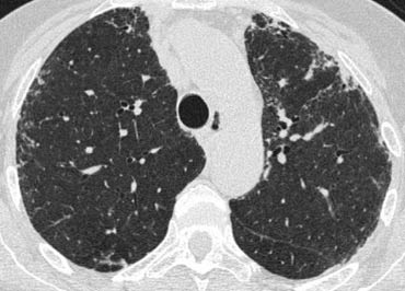
Tree-in-bud refers to a pattern seen on thin-section chest CT in which centrilobular bronchial dilatation and filling by mucus pus or fluid resembles a budding tree Usually somewhat nodular in appearance the tree-in-bud pattern is generally most pronounced in the lung periphery and associated with abnormalities of the larger airways. It represents dilated and impacted mucus or pus-filled centrilobular bronchioles.
The Radiology Assistant Hrct Basic Interpretation
The Radiology Assistant Hrct Basic Interpretation

ボード Radiography のピン
The Radiology Assistant Hrct Common Diagnoses
2
2
Tree In Bud Sign Lung Radiology Reference Article Radiopaedia Org
The Radiology Assistant Hrct Basic Interpretation

Pin By Arlene Boyden On Excalibur Healthcare S Imaging Teleradiology Pins Radiology Radiography Medical Knowledge

Tree In Bud Pattern
2

Learningradiology Lung Abscess Pulmonary Lunges Pulmonary X Ray
The Radiology Assistant Hrct Common Diagnoses
Chronic Airspace Disease Review Of The Causes And Key Computed Tomography Findings
Mycobacterium Avium Complex Mac Mycobacterium Avium Intracellulare Mai Workup Approach Considerations Ats Idsa Diagnostic Guidelines Acid Fast Bacillus Stains And Cultures

Imaging Patterns Of Pneumonia Sciencedirect
The Radiology Assistant Cystic Lung Cancer

Fig 7 Tree In Bud Sign Chest Ct Shows Tree In Bud Images Schematic Drawings And Corresponding Picture Refe Radiology Radiology Imaging Medical Radiography
The Radiology Assistant Hrct Basic Interpretation